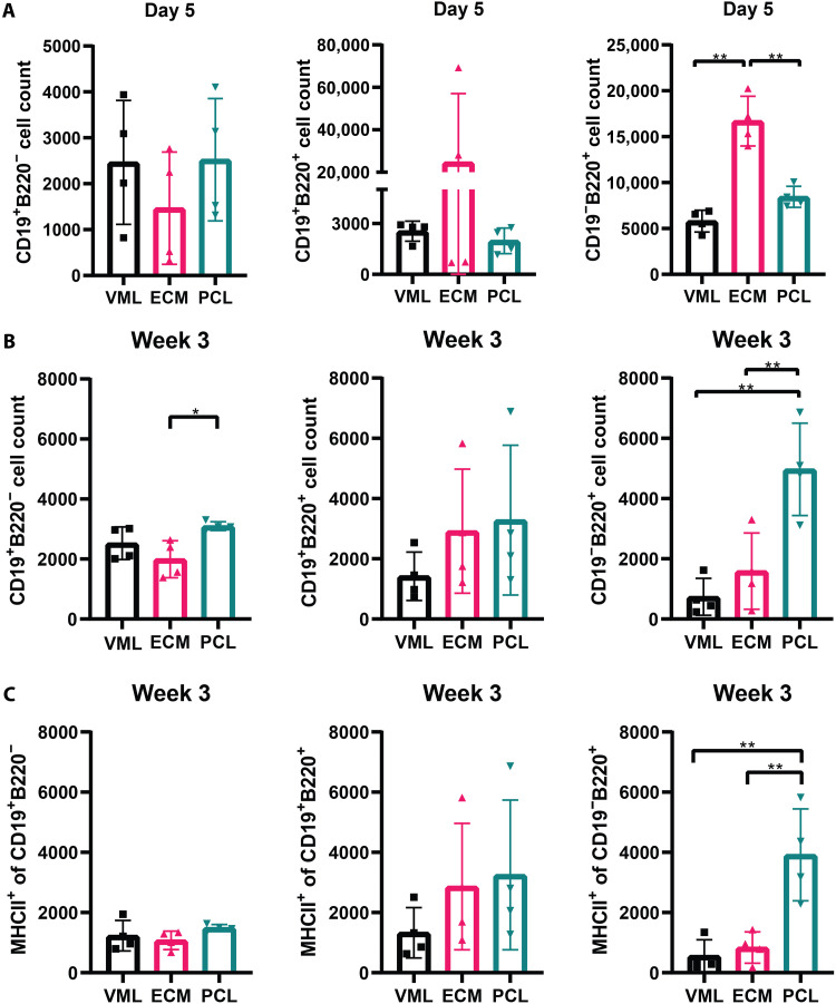
Fig. 3. PCL induces B cell antigen presentation in the quad tissue.
(A) Flow cytometry counts of B cell subsets: CD19+B220−, CD19−B220+, and CD19+B220+ in the quad tissue at day 5 after injury. (B) MHCII+B220+ cell population in quadricep tissue at day 5 after injury. (C) Flow cytometry counts of B cell subsets: CD19+B220−, CD19−B220+, and CD19+B220+ in the quad tissue at week 3 after injury. Data from CD19−B220+ are also used in Fig. 1A. Data are means ± SD, n = 4, two-way ANOVA with subsequent multiple comparison testing [(B and C): *P < 0.05, **P < 0.01]. VML indicates injury with saline, ECM indicates injury with ECM, and PCL indicates injury with PCL.

