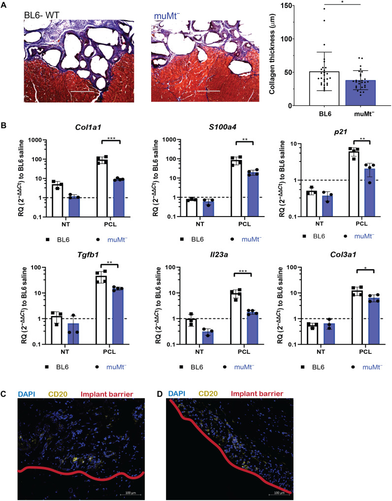
Fig. 4. muMt− reduces PCL-mediated fibrosis.
(A) Histological staining of 6-week PCL implants in BL6-WT and muMt− with Masson’s trichrome for collagen fibrils. Scale bar, 400 μm. (B) Gene expression analysis of fibrosis-related genes including Col1a1, S100a4, p21, Tgfb1, Il23a, Col3a1 via qRT-PCR. (C) Immunofluorescence staining of CD20+ cells (yellow) and the implant barrier (red) in human tissue samples surrounding breast capsule implants of acellular adipose tissue (AAT) and (D) of silicone. Scale bar, 100 μm. Data are means ± SD, n = 3 for NT controls, n = 4 for PCL biomaterial, two-way ANOVA with subsequent multiple comparison testing [(C): ***P < 0.001, **P < 0.01, *P < 0.05]. muMt− indicates mice that lack mature B cells, BL6 indicates C57BL6 mice, VML indicates injury with saline, ECM indicates injury with ECM, and PCL indicates injury with PCL.

