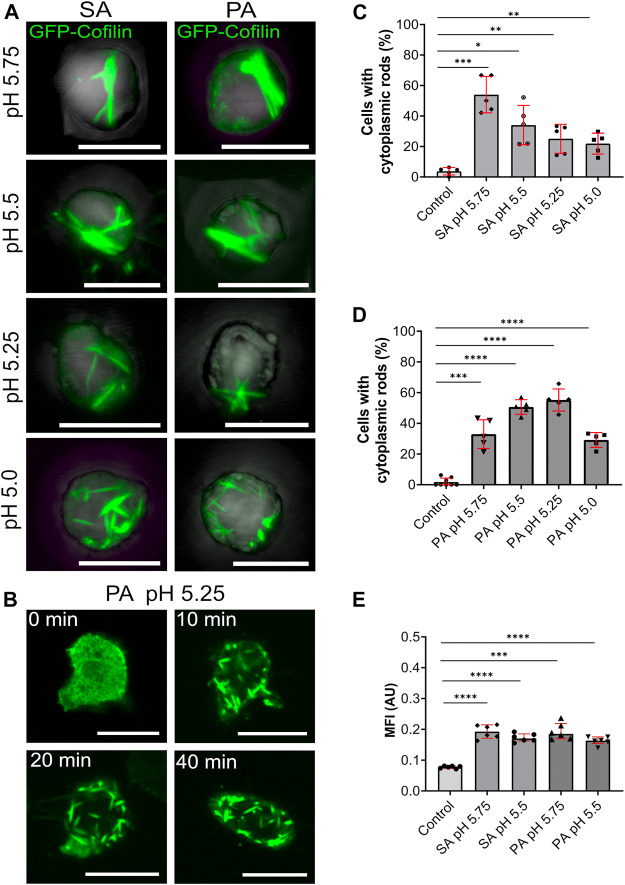
FIGURE 4.
Cytoplasmic actin-cofilin rods are induced by lowering the intracellular pH. (A) 3D projection images of GFP-cofilin rods (green) formed by GFP-cofilin expressing cells cultivated for 2 h in medium adjusted to lower pH values with sorbic acid (SA) or propionic acid (PA) (pH 5.75, 5.5, 5.25 and 5.0). Scale bars are 10 µm. (B) Time lapse images of GFP-cofilin expressing cells placed in medium adjusted to pH 5.25 with PA. After 10 min, GFP-cofilin rods (green) start to assemble in the cytoplasm. Scale bars, 10 µm. (C,D) Percentage of cells with GFP-cofilin rods in medium adjusted with SA (C) or PA (D) to the indicated pH values. n = 5 experiments. (E) Ratiometric pH measurements to determine acidification of the intracellular pH. Dictyostelium cells expressing pHluorin2 cells were incubated with medium adjusted with either SA or PA to pH 5.75 and 5.5. An increase of MFI indicates the lowering of the intracellular pH. n = 6 experiments per treatment. More than 20 cells were analyzed for each experiment and treatment. Error bars are median with interquartile range (red). Samples were considered significant when p ≤ 0.05. Welch´s t-test was applied.

