Figure 2.
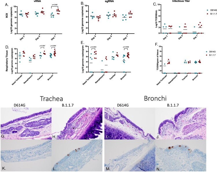
Virus load, pathology and virus antigen in the respiratory tract. AGMs were infected with either the D614G or B.1.1.7 SARS-CoV-2 variant intranasally utilizing the Nasal Mucosal Atomization Device. Bronchial cytology brush (BCB) samples were collected on days 3, 5 and 7 post-infection and samples were analyzed for vRNA, sgRNA and infectious virus. (A-C) A significant difference in vRNA collected in the BCBs was detected 7 days-post-infection (A, multiple unpaired t-test, p-value <0.05). Animals were euthanized on day 7 post-infection and respiratory tissues were collected for analyses for vRNA, sgRNA and infectious virus (D-F). vRNA was significantly different in the trachea and bronchi (D, multiple unpaired t-test, p-value <0.05). sgRNA differed significantly in the nasal turbinate and bronchi (E, multiple unpaired t-test, p-values <0.05). Infectious virus did not differ significantly in any tissue Infectious virus did not differ significantly in any tissue (F, multiple unpaired t-test were used to compare tissues between groups). Pathology and immunoreactivity in the trachea and bronchi (G-N). Normal trachea found in the D614G (G) vs. trachea with cellular infiltrates, hemorrhage (*) and fibrin (arrow) found in the submucosa in the B.1.1.7 (H). Immunoreactivity in the trachea of D614G vs B.1.1.7. (K, L, respectively) Normal bronchi found in the D614G (I) vs bronchi with inflammation and cellular infiltrates found in the B.1.1.7. (J) Immunoreactivity in bronchi of the D614G vs B.1.1.7 (M,N) (H&E and IHC magnifications (G_N) = 100x).

