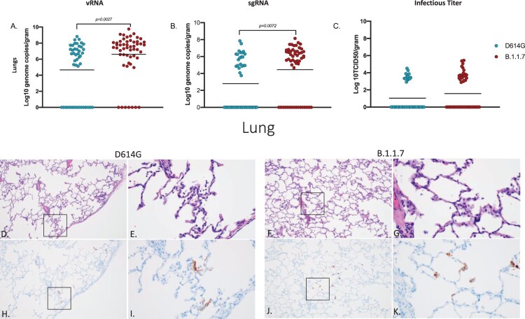
Figure 3.
Virus load, pathology and immunoreactivity in the lungs. Animals were euthanized on day 7 post-infection and a section from each lung lobe was collected and analyzed for vRNA, sgRNA and infectious virus. Results of each assay were combined to look at the lungs in total (A-C). Significant differences were detected in lungs in both vRNA and sgRNA (D, multiple unpaired t-test, p-value <0.05 and E, multiple unpaired t-test, p-value <0.05) but not in infectious virus (C). Multiple unpaired t-tests were used to compare the two groups for statistical significance. Pathology and immunoreactivity in the lungs (D-K). Minimally thickened and inflamed alveolar septa with multifocal pneumocyte immunoreactivity in the D614G and B.1.1.7 samples (H&E (D,F) and IHC (H,J) = 100x; H&E (E,G) and IHC I,K) = 400x.).

