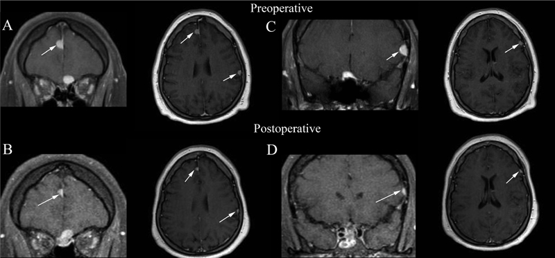
Fig. 3.

Coronal and axial T1-weighted magnetic resonance imaging slices 10 months postoperative and 4 months postmegestrol cessation demonstrating interval decrease in size of multiple meningiomas, including: right anterior falx ( A ) 11 mm, now ( B ) 5 × 4 × 8 mm, left parietal calvarial ( A ) 7 × 10 mm, now ( B ) 3 × 5 mm and lateral frontal calvarial based lesion ( C ) 8 × 11 mm, and now ( D ) 7 × 4 mm.
