Figure 1.

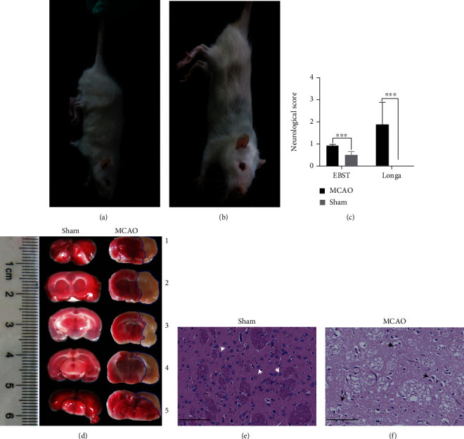
Construction and evaluation of MCAO model for brain injury following ischemia and reperfusion. (a) MCAO rat behavior observations. Longa rating of 1 confirmed the success of our MOCA rat model. Behavior manifested as mild neurological deficit symptoms, whereby forelimbs could not be fully extended. (b) Sham rat behavior observations. Forelimbs could be naturally extended without neurobehavioral deficits. (c) Longa and EBST scores. Data are expressed as the means ± SEMs and compared using Student's t-test (∗∗∗P < 0.001 versus sham, n = 5 mice per group with triplicate biological replicates). (d) TTC-stained brain sections. Ischemic area stained white (lined with blue) and the intact area stained red. Left: representative coronal brain sections of the sham group; right: MCAO group at 2 h MCAO followed by 24 h of reperfusion. (e, f) H&E-stained brain slices of sham and MCAO groups, respectively (scale bar = 100 μm). Neurons in (e) were tightly aligned with moderate and distinct nucleoli. The white arrows indicate undamaged nerve cells with intact structural morphology. Owing to the presence of vacuolation and mesenchymal edema, staining of the infarcted area was lighter than that of the sham group. The black arrows indicate pathological abnormalities with loosely arranged neurons, pyknotic nuclei, nuclear lysis, and vacuole-like degeneration.
