. 2021 Nov 11;10:e72798. doi: 10.7554/eLife.72798

Figure 1. UCH37–RPN13C preferentially cleaves branched polyUb chains.

Branched (A) or linear (B) Ub3 substrates (5 μM) with the indicated Ub–Ub linkages were incubated for 1 hr at 37°C with 1, 5, or 10 μM His-TEV-UCH37, with or without the addition of equimolar RPN13C. Reactions were analyzed by sodium dodecyl sulphate-polyacrylamide gel electrophoresis (SDS–PAGE) and Coomassie staining. (C) Quantification of the Ub2 and Ub1 products from (A) and (B). For linear Ub3 substrates, results from incubations with 10 μM enzyme are shown; for branched Ub3 substrates, results from 1 μM enzyme are shown. (D) Wild-type (WT), M148A F149A (AA), or M148D F149D (DD) UCH37, alone or with the addition of equimolar of RPN13C, were incubated with Ub3 substrates for 2 hr at 37°C. Left, reactions contained 0.5 μM enzymes and 10 μM branched substrates. Right, reactions contained 10 μM enzymes and 5 μM linear Ub3. (E) Quantification from (D), plotted as the ratio of Ub1 produced in the presence over absence of RPN13C. Mean ± standard deviation (SD) from two independent replicates are shown.

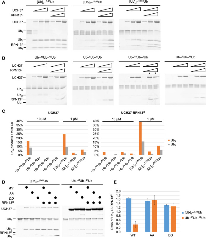
Figure 1—source data 1. Uncropped gels in Figure 1.

Figure 1.

Figure 1—figure supplement 1. UCH37–RPN13C preferentially cleaves branched polyUb chains.

Figure 1—figure supplement 1.

(A) Notation used to describe architectures of polyUb chains used in this paper (adopted from Varadan et al., 2005). Note that we use the term ‘linear’ to refer to nonbranched Ub chains, which include but are not exclusively M1-linked Ub chains. (B) UCH37 exclusively cleaves the K48 linkage in a branched chain. Ub3 substrates were incubated with the indicated DUB (i.e., UCH37–RPN13C complex or OTUB1) and the reaction products were detected using either the K6 linkage-specific anti-Ub affimer (Michel et al., 2017) or K48 linkage-specific anti-Ub antibody. In Lane 5, OTUB1 was added after UCH37–RPN13C had completely hydrolyzed the K6/K48-branched Ub3, thereby producing K6-linked Ub2 that was refractory to OTUB1. (C) Crystal structure of the UCH37–RPN13C–Ub complex (PDB code: 4WLR). Part of the active site crossover loop (ASCL) is disordered and is represented as a dotted line. UCH37 ASCL residues M148 F149 contact RPN13C and are shown in gray. UCH37 residues E34, W36, and I216 contact the hydrophobic patch on Ub and are shown in orange. UCH37 catalytic-site residue C88 is shown in red. (D) Quantification from Figure 1D, plotted as the ratio of Ub2 in the presence over absence of RPN13C. Mean ± standard deviation (SD) from two independent replicates are shown.
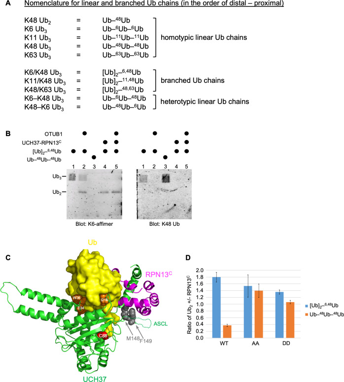
Figure 1—figure supplement 1—source data 1. Raw western data in supplement 1B.