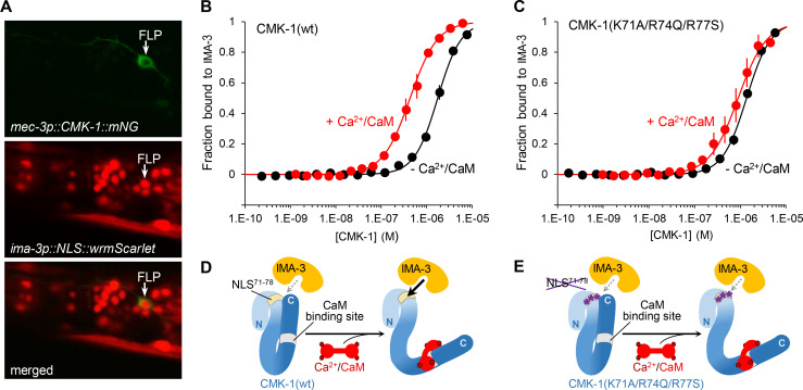
Figure 4. Ca2+/CaM binding to CMK-1 enhances the affinity for IMA-3 importin-α in a NLS71-78-dependent manner.
(A) ima-3 expression analysis with a transcriptional reporter. Representative confocal micrographs showing fluorescence signals in the head of an adult [mec-3p::CMK-1::mNG; ima-3p::NLS::wrmScarlet] transgenic animal. FLP cytoplasm is labeled in green (top). The nuclei of ima-3-expressing cells are labeled in red (middle). FLP is among the cells expressing ima-3p (bottom). (B) Binding curves for the CMK-1(wt)/IMA-3 interaction in the presence (red curve) or the absence (black curve) of Ca2+/CaM, as determined by MicroScale Thermophoresis (see Materials and methods). Data as average of three replicates (± SEM). (C) Same analysis as in (B), but with CMK-1 (K71A/R74Q/R77S) mutant. Kd derived from fitting curves are reported in Table 1. (D, E) Schematic protein interaction models illustrating the increased affinity for IMA-3 when Ca2+/CaM binds CMK-1(wt) (D) and the impact of NLS71-78 mutations, which prevent CaM-dependent IMA-3 affinity increase.

