FIGURE 3.
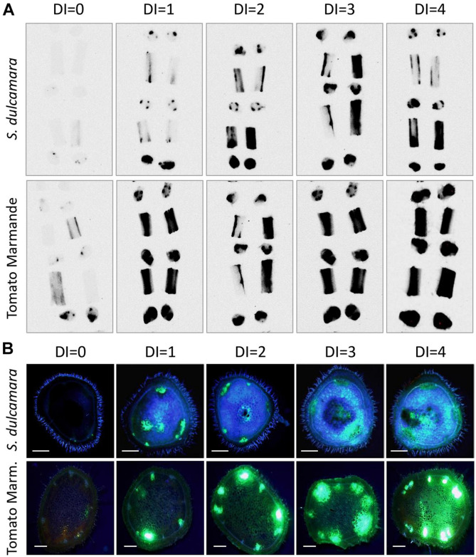
R. solanacearum distribution in stems of inoculated S. dulcamara and tomato cv. Marmande plants. (A) Representative luminescence imaging photographs at different wilting stages (Disease index 0–4) of stem sections from S. dulcamara (top panel) and tomato (bottom panel) plants stem-inoculated with luminescent R. solanacearum. Luminescent bacteria are detected as dark areas in transversal and longitudinal sections of plant internodes 1–4 organised bottom to top. Inoculation points are indicated by an arrow. (B) Representative fluorescence microscopy images of stem sections from S. dulcamara (top panel) and tomato (bottom panel) plants stem-inoculated with an R. solanacearum strain constitutively expressing GFP. Inoculations were performed at the base of the first true leaf and transversal sections obtained in the first internode, 2 cm above the inoculation point. Scale bars indicate 0.5 mm.

