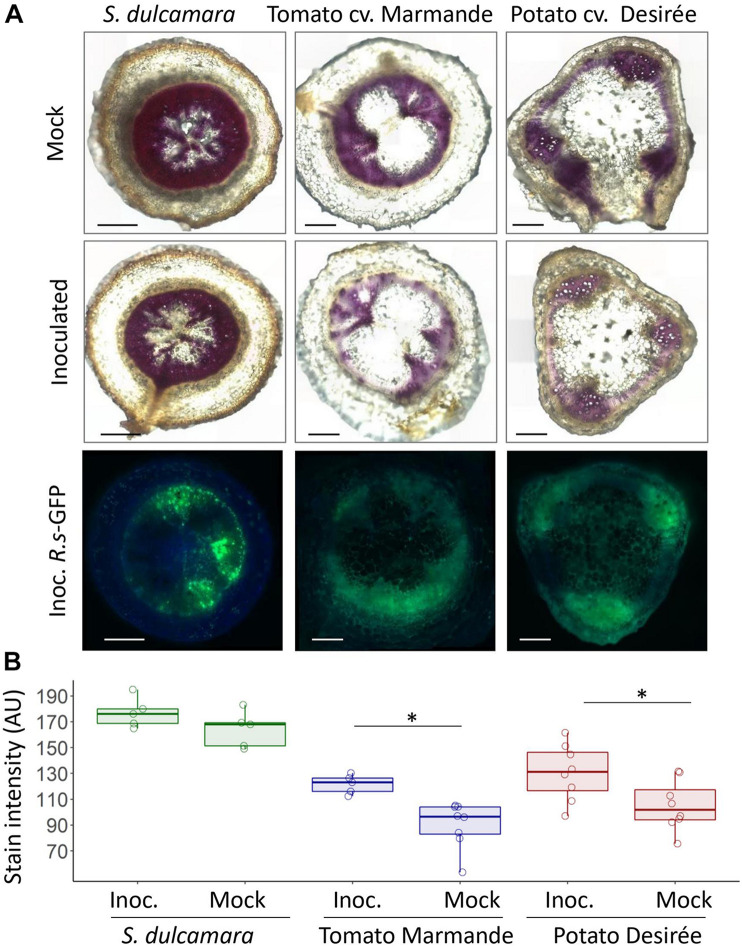
FIGURE 5.
Lignification of S. dulcamara, Tomato cv. Marmande and S. tuberosum cv. Desirée tissues upon R. solanacearum infection. (A) Representative composed images of S. dulcamara, tomato, and potato taproot transversal sections obtained 9 days after inoculation with R. solanacearum-GFP or mock treatment. First and second row: microscope bright-field images after lignin staining with phloroglucinol HCl (magenta colouration). Third row: fluorescence microscopy images after inoculation to assess the extent of bacterial colonisation. Images were obtained using a Leica DM6 microscope. Scale bars indicate 0.5 mm. (B) Quantification of the phloroglucinol HCL stain -indicative of lignin content- in the vascular area from the images shown in A performed with the ImageJ software. ∗indicates statistical differences (p-value < 0.05; T-student significant test α = 0.05).

