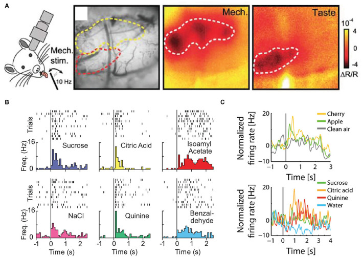
Figure 2.
(A) In vivo intrinsic optical imaging of the oral-somatosensory cortex and gustatory cortex; note that the cortical region responding to taste stimuli is located ventral to the area activated by the tactile stimulation of the tongue. (B) The single-unit activity of a neuron in the gustatory cortex in response to the intraoral delivery of individual taste stimuli (sucrose, NaCl, citric acid, and quinine) and individual odors dissolved in water (isoamyl acetate and benzaldehyde). (C) The normalized activity of a neuron in the posterior piriform cortex in response to orthonasally presented odors (apple, cherry, and clean air) and intraorally delivered taste stimuli (sucrose, citric acid, quinine, and water). Panels adapted from (A) Copyright (2007) Society for Neuroscience (Accolla et al., 2007), (B) (Samuelsen and Fontanini, 2017), and (C) (Maier et al., 2012).

