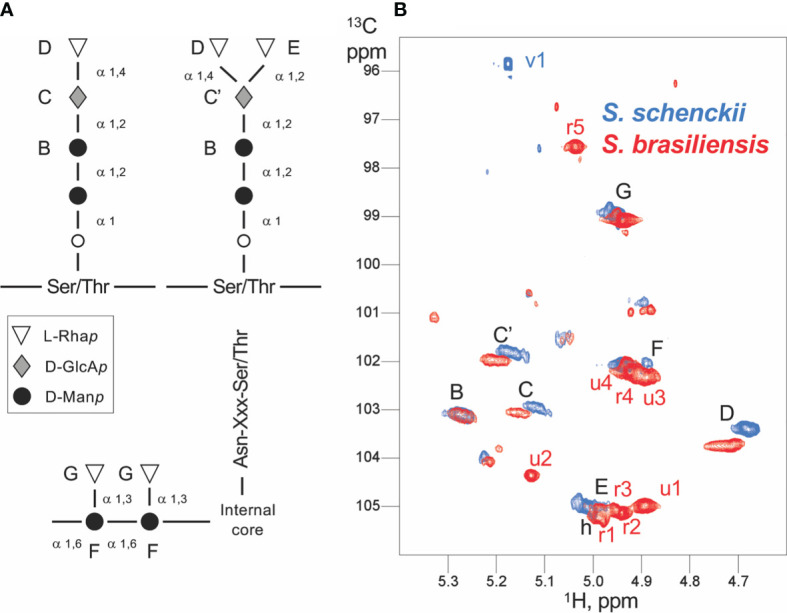
Figure 8.

Scheme of the Sporothrix peptidorhamnomannan (PRM) organization and NMR assignments. (A) The generally known structures of the O-linked (top) and N-linked rhamnomannan (bottom) are shown. The inter-residue linkage types are displayed. The capital letters from B to G denote the assignments obtained by NMR, and the corresponding signals are displayed in panel (B). (B) Anomeric region of the 1H-13C HSQC spectra of Sporothrix schenckii (blue) and Sporothrix brasiliensis (red) crude PRMs. Each signal arises from the anomeric 1H-13C group of a distinct polysaccharide unit. The signals common to both species (and thus common residues) are labeled in black, whereas the signals unique to a species are labeled in the corresponding color. Capital letters denote signals from assigned residues; the corresponding residues are labeled in the PRM organization scheme (A) . The mannose O-linked to Ser/Thr could not be assigned. Whereas S. schenckii only shows a single residue absent (v1) in S. brasiliensis, the later displays at least nine unique residues with five identified rhamnose units (r1 to r5) and four yet unidentified units (u1–u4).
