
Figure 1.
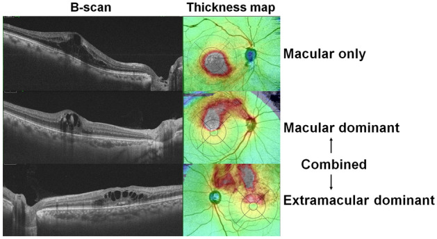
Pattern of retinal edema at baseline in eyes with macular edema secondary to branch retinal vein occlusion (BRVO). All horizontal B-scans across the fovea shows cystoid macular edema. However, the extent and topographic features of retinal edema vary from edema exclusively in the macula (A) and combined extramacular and macular edema (B, C). Depending on the dominant area of edema (white or red areas), eyes with combined edema were separated into macular dominant (B) and extramacular dominant edema (C).