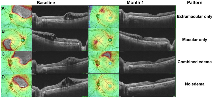
Figure 2.
Pattern of retinal edema 1 month following anti-vascular endothelial growth factor (VEGF) therapy for macular edema (ME) due to branch retinal vein occlusion (BRVO). Four patterns, (extramacular only [A], macular only [B], combined edema [C], and no edema [D]) are noted from the widefield retinal thickness maps. The areas of residual edema are mostly wedge-shaped and located between the fovea and site of vascular occlusion.

