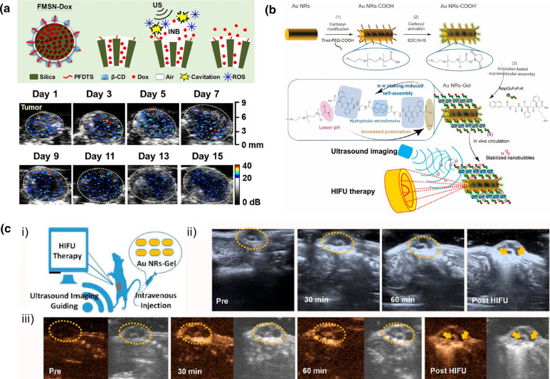
Fig. 7.
Examples of GSNs used for in vivo ultrasound imaging. a Schematic showing drug loaded hydrophobic MSNs coated with β-CD (above) and in vivo ultrasound contrast enhancement as a result of cavitation generation by GSNs over 15 days.
Adapted from reference [40], Copyright 2020, Elsevier. b Schematic showing the preparation of peptide hydrogel coated gold nanorods and nanobubble stabilization by them. c Ultrasound contrast enhancement by intravenously injected peptide hydrogel coated gold nanorods at different time points after nanoparticle injection and after HIFU treatment. Adapted from reference [84], Copyright 2019, American Chemical Society

