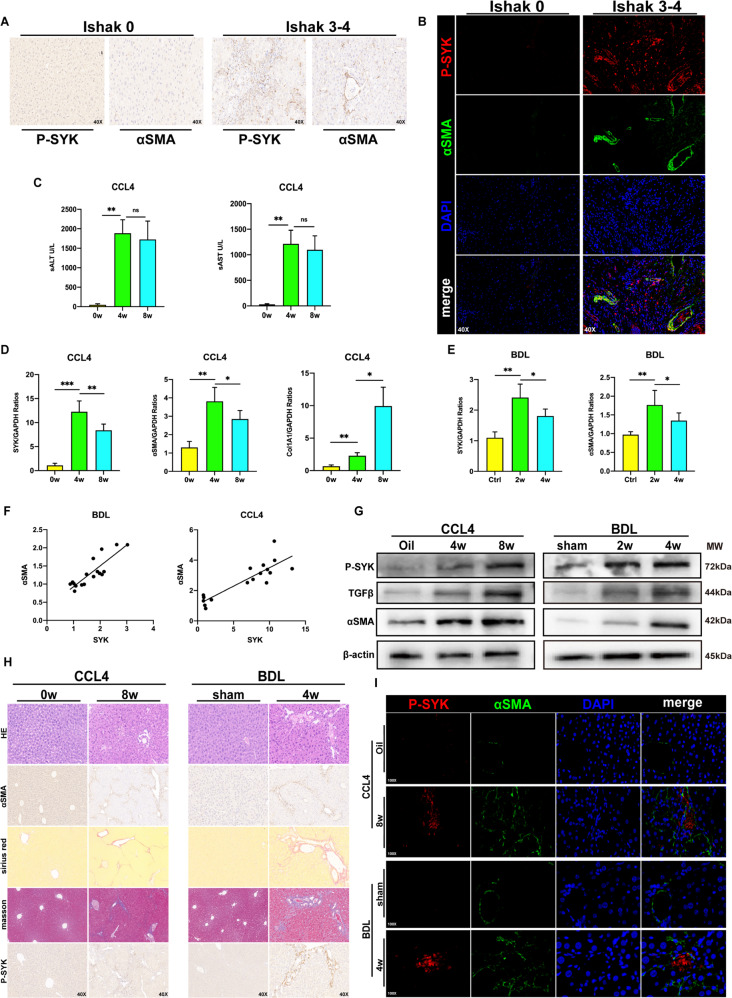
Fig. 1. p-SYK is essential for human and mice liver fibrosis.
A Immunohistochemical staining of p-SYK and α-SMA in liver tissues of fibrosis patients with different ishak grades (Ishak0, Ishak3–4) (magnification 40X). B Confocal immunofluorescence staining of p-SYK and α-SMA in liver tissues of fibrosis patients with different ishak grades (Ishak0, Ishak3–4) (magnification 40X), DAPI as nuclear localization. C Serum ALT and AST levels in mice injected with CCL4 for 4 and 8 weeks, oil as a control. D Assessed the expression of SYK, α-SMA, and col1A1 in the liver of mice treated with CCL4 for 4 and 8 weeks were by qPCR, and oil was used as a control. E The expression of SYK and α-SMA in the liver of mice 2 and 4 weeks after BDL surgery were determined by qPCR, and sham was used as a control. F Correlation analysis between the expression of SYK and α-SMA in CCL4 and BDL models. G Western Blot was used to analyze the protein level of p-SYK, TGF-β and α-SMA in the liver of mice injected with CCL4 for 4 or 8 weeks and mice 2 or 4 weeks after BDL, oil and sham were used as controls, respectively. H HE, Sirius red, masson staining and immunohistochemistry of α-SMA and p-SYK were performed on the livers of mice given CCL4 for 8 weeks and mice 4 weeks after BDL, respectively. Oil and sham were used as controls. I Confocal immunofluorescence staining of p-SYK and α-SMA were performed on the livers of a mouse injected with CCL4 for 8 weeks and mice 4 weeks after BDL, respectively, with DAPI as the nuclear localization.

