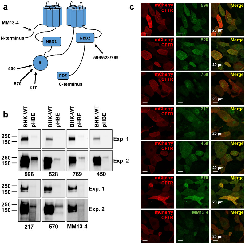
Figure 1.
Immunodetection of CFTR. (a) Cartoon showing locations of epitopes for the seven anti-CFTR antibodies used in this study. (b) Immunoblots of lysates after SDS-PAGE probed for CFTR using each antibody. BHK-WT cells were stably transfected with CFTR. Well differentiated primary bronchial epithelial (HBE) cells expressed endogenous CFTR. Protein (BHK-10 µg, HBE-40 µg) was detected after short and long exposures (Exp.1 and 2, respectively). Blots were cropped. (c) Confocal images of HBEs at the apical membrane after transduction with adenoviral mCherry-WTCFTR. Cells were cultured at the ALI for 10–15 days and immunostained on intact membrane support.

