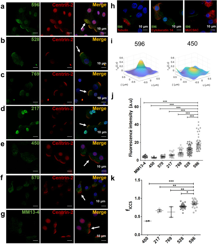
Figure 2.
The bases of cilia are immunostained using CFTR antibodies 596, 528, 769 and 217, but not 450, 570 and MM13-4. (a–g) Confocal images of well differentiated HBEs co-immunostained with CFTR antibodies 596, 528, 769, 217, 450, 570 or MM13-4 and the ciliated cell marker centrin-2. Arrows indicate ciliated cells. (h) Confocal images of cells coimmunostained with 596 and antibodies against either tubulin (left, ciliated cell marker), cytokeratin 14 (middle, basal cell marker) or MUC5AC (right, goblet cell marker). (i) Representative spatial cross-correlation functions calculated via ICCS analysis between centrin-2 and CFTR immunofluorescence using 596 (left) or 450 (right) measured from image sub-regions of interest (64 × 64 pixels). (j) Average apical immunofluorescence signals with different CFTR antibodies. Intensities were normalized to the excitation power (mean ± SD, n = 12–57, ***p < 0.0001, one-way ANOVA). (k) Colocalized fraction fICCS extracted from ICCS analysis (mean ± SD, n = 2–28, 450 vs 596, ***p < 0.0001; 217 vs 596, **p = 0.005; 769 vs 596, **p = 0.0059; 528 vs 596, *p = 0.0313), which indicates the arithmetic mean of the fractions of CFTR and centrin-2 antibody signals that interact.

