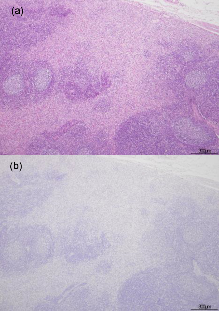
Fig. 2.

Hematoxylin & eosin and immunohistochemical staining for African swine fever phosphoprotein p30 in lymph nodes. Marked fibrosis predominantly presented in the sinuses, especially in the subcapsular area of case no. 2 (a). Antigen-labeled cell was not present in the lesion of case no. 2 (b).
