. 2021 Sep 15;3(6):e210031. doi: 10.1148/ryai.2021210031

Figure 2:

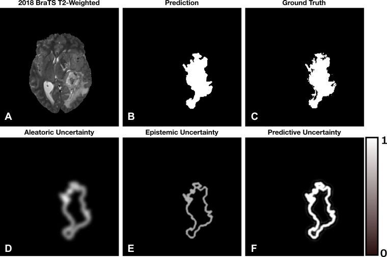
A simulated example of the aleatoric, epistemic, and predictive uncertainties for a segmentation task. Brighter pixels indicate larger uncertainty with range 0–1. (A) Axial T2-weighted MRI section from the 2018 Medical Image Computing and Computer Assisted Intervention Society Multimodal Brain Tumor Image Segmentation dataset (16,17). (B) The hypothetical segmentation output. (C) The ground truth segmentation. (D) Aleatoric uncertainty localized to the boundaries of the segmentation. (E) Epistemic uncertainty localized to the boundary of the segmentation with less ambiguity compared with the aleatoric uncertainty. (F) Predictive uncertainty, which is the addition of (D) and (E). We notice that the model is confident within the interior of the segmented lesion and less so at the boundary.

A simulated example of the aleatoric, epistemic, and predictive uncertainties for a segmentation task. Brighter pixels indicate larger uncertainty with range 0–1. (A) Axial T2-weighted MRI section from the 2018 Medical Image Computing and Computer Assisted Intervention Society Multimodal Brain Tumor Image Segmentation dataset (16,17). (B) The hypothetical segmentation output. (C) The ground truth segmentation. (D) Aleatoric uncertainty localized to the boundaries of the segmentation. (E) Epistemic uncertainty localized to the boundary of the segmentation with less ambiguity compared with the aleatoric uncertainty. (F) Predictive uncertainty, which is the addition of (D) and (E). We notice that the model is confident within the interior of the segmented lesion and less so at the boundary.