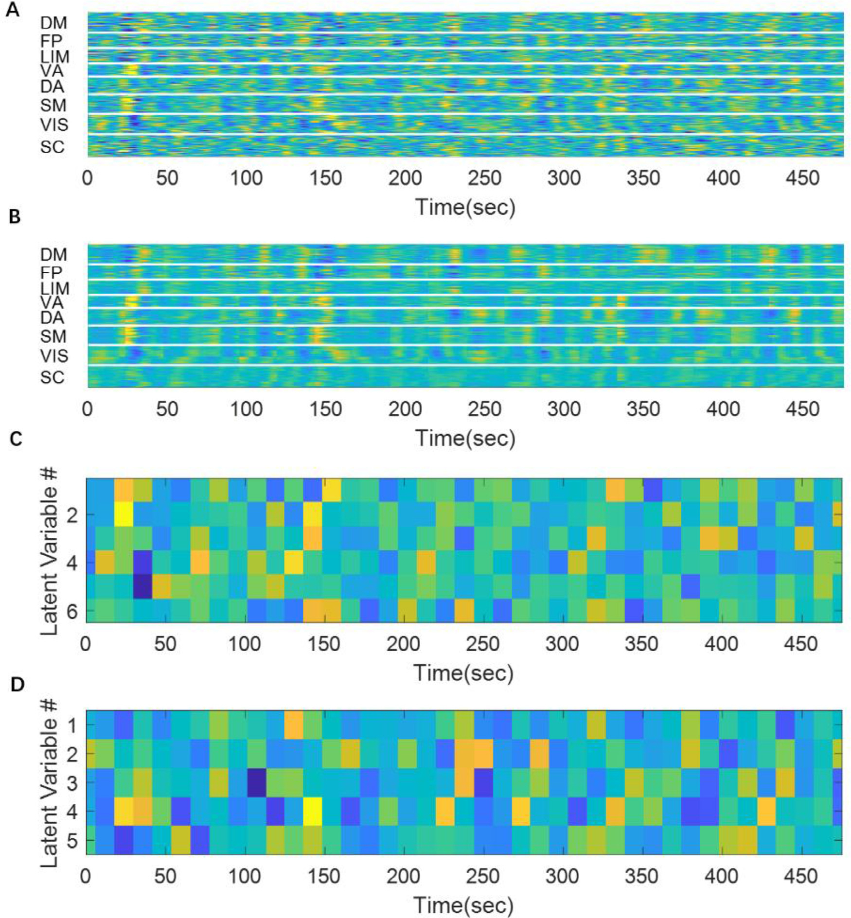
Fig. 6. A fMRI segment can be encoded as a 32-dimensional code.

Panel A shows 20 concatenated original rs-fMRI segments. Panel B shows the reconstructed rs-fMRI segments. It can be seen that the reconstruction matches fairly well with the original signal although there are some abrupt changes at the edge of each segment (that were concatenated together). Panel C and D show the values of latent variables in cluster1 and cluster 2, respectively. The remaining 4 clusters were not shown for display purposes.
