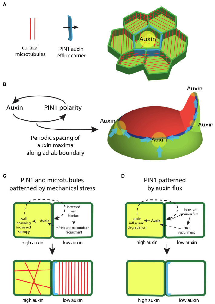
Figure 3.
The regulation of auxin transport within the shoot epidermis. (A) In the shoot epidermis, the auxin efflux carrier PIN-FORMED1 (PIN1; blue) forms supracellular polarity convergence patterns that concentrate auxin locally. PIN1 localization within cells correlates with the orientation of interphase microtubule arrays (red), suggesting a role for mechanical stresses in regulating PIN1. (B) Auxin influences the polarity of PIN1 in a feedback loop. Modeling indicates this feedback is sufficient to generate a periodic spacing between PIN1 polarity convergence patterns (blue) and thus auxin maxima (yellow) along the ad-ab boundary (dark blue) located between adaxial (red) and abaxial (green) tissues. (C) Illustration depicting how differential auxin concentrations might regulate PIN1 and microtubule localization via changes to mechanical stress. High levels of auxin (yellow) in one cell triggers cell wall loosening. This leads to higher tension levels within the cell wall of an adjacent cell, which acts to polarize PIN1 (blue) and orient cortical microtubules (red). (D) Illustration depicting how high levels of auxin might regulate PIN1 via changes in auxin flux. High levels of auxin (yellow) triggers the expression of the auxin influx carrier as well as auxin degradation. This promotes an increase in diffusive auxin into the cell from adjacent cells. Increased auxin efflux in the adjacent cells promotes a corresponding PIN1 polarity (blue).

