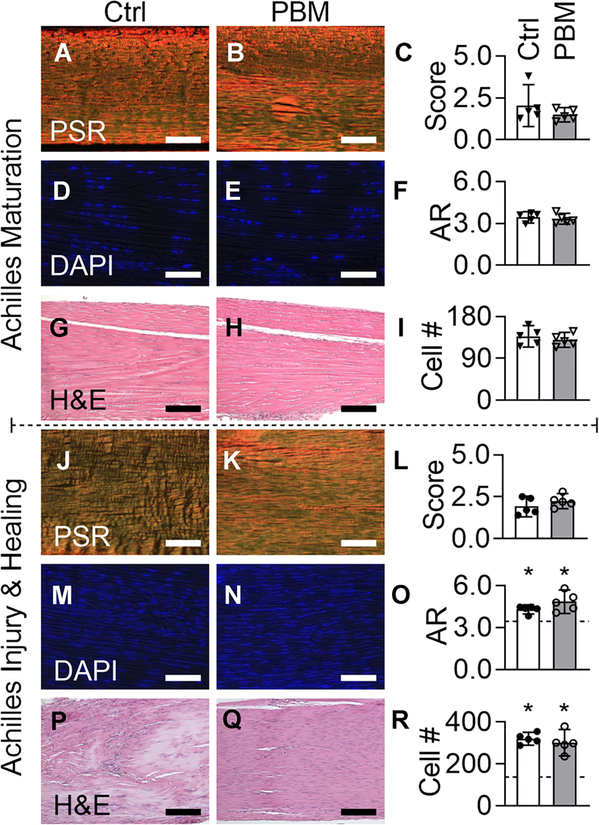
FIGURE 2.
During maturation and adult injury and healing, daily doses of PBM therapy did not affect the structural or cellular properties of the mouse Achilles tendon. (A-C, maturation; J-L, healing) Collagen alignment, (D-F, maturation; M-O, healing) nuclear aspect ratio (AR) and (I, maturation; R, healing) cell number, and (G and H, maturation; P and Q healing) fibrosis were observed for control (Ctrl, white bars) and PBM (gray bars) treated tendons using Picrosirius Red (PSR), DAPI, and Hematoxylin and Eosin (H&E) stained sections, respectively. O and R, Dashed line is the mean of maturation controls and asterisks represent a significant difference from maturation controls (P < .05). Scale bar = 75 μm for panels A and B, G and H, P and Q, and 40 μm for panels D and E and J-N. Maturation and healing data points are downward arrowheads and circles, respectively. Data are presented as mean ± standard deviation. Ctrl, control; DAPI, 4',6-diamidino-2-phenylindole; PBM, photobiomodulation

