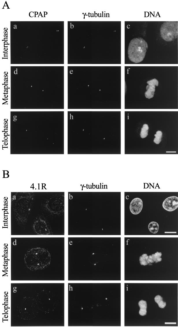
FIG. 6.
(A) Confocal microscopy analysis of the subcellular localization of human CPAP and γ-tubulin in various cell dividing stages. SiHa cells were triply stained with affinity-purified anti-CPAP antibody, a γ-tubulin MAb, and a DNA-specific dye, propidium iodide. (B) Triple immunostaining of SiHa cells with anti-C-4.1R and γ-tubulin antibodies. DNA was stained by propidium iodide. Each image represents a confocal optic section, and only the sections that revealed the centrosomal localization were selected and composed in panel B. By adjusting the focus, 4.1R was seen in the nucleus as well as in the cytosol (which are located at different focal planes). Bars, 10 μm.

