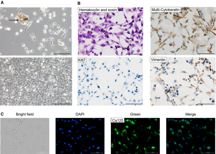
Fig. 2.

Morphology and identification of ascites‐derived epithelial ovarian cancer (EOC) cells. (A) Representative phase‐contrast images of EOC cells. In the upper image: cells after initial isolation, grape‐like cluster (arrow), and single EOC cells. In the lower image: a confluent monolayer of EOC cells expanded in culture. Note the typical epithelial cobblestone morphology and tight cell‐to‐cell junction. (B) Subsequent cytological analysis. Immunochemistry of EOC cells showed expression of epithelial marker multicytokeratin, mesenchymal marker, vimentin, and cell proliferation marker Ki67. (C) Immunofluorescence staining of EOC cells revealed the expression of the Mucin‐16 tumor marker (CA125). Brightfield, nucleus staining with blu DAPI, Mucin‐16 marker staining with green Alexa Fluor® 488, and merge image (DAPI+ Alexa Fluor® 488) are reported. Bar = 100 μm. For each set of analyses, representative results out of three independent experiments are shown.
