Table 2.
Detailed information regarding athetes with post-COVID-19 with myocardial or pericardial alterations on cardiac MRI
| Athlete no | Sex | Symptoms | Findings on other exams | Time to CMR after positive test results (days) | hsTnT recorded prior to CMR (ng/L) | CMR findings | Pathological alteration | Certainty of cardiac involvement | Clinical outcome (6 months) |
| 1. | Male | Moderate
|
Troponin: elevated (hsTnT: 18 ng/L, normal: <14 ng/L) 12-lead ECG: minor repolarisation. alteration Holter ECG: sinus tachycardia (1 hour) Echocardiography: slightly dilated RV Exercise test (4 months after COVID-19 infection): normal |
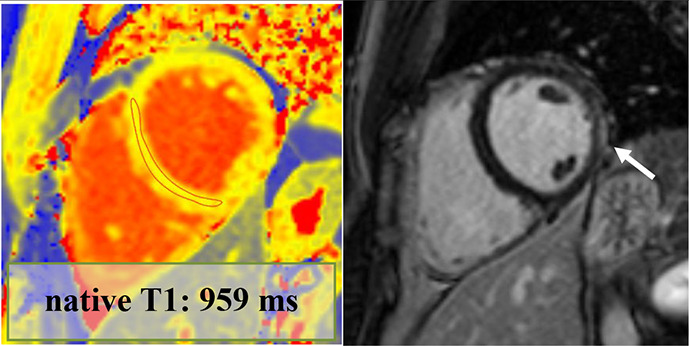
70 | 18 | LVEF: 52% GLS: −18% Septal native T1: normal Septal native T2: normal Pathological LGE/pattern: yes—lateral subepicardial T1 and T2 mapping value in the area corresponding with the LGE: 1016 and 50 ms—mildly elevated |
|
Definite | Returned to sport, no persistent cardiac complaints at follow-up |
| 2. | Male | Moderate
|
Troponin: elevated (hs troponin I: 198 ng/L, normal: <45 ng/L) 12-lead ECG: minor repol. alteration Holter ECG: normal Echocardiography: normal Exercise test (3 months after COVID-19 infection): normal |
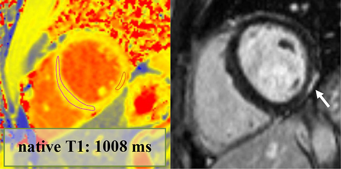
74 | NA | LVEF: 58% GLS: −18% Septal native T1: elevated Septal native T2: normal Pathological LGE/pattern: Yes—lateral subepicardial T1 and T2 mapping value in the area corresponding with the LGE: 1065 and 53 ms—elevated |
|
Definite | Returned to sport, no persistent cardiac complaints at follow-up |
| 3. | Male | Moderate
|
Troponin: normal 12-lead ECG: RBBB (previously reported) Holter ECG: not performed Echocardiography: normal Exercise test (5 months after COVID-19 infection): normal |
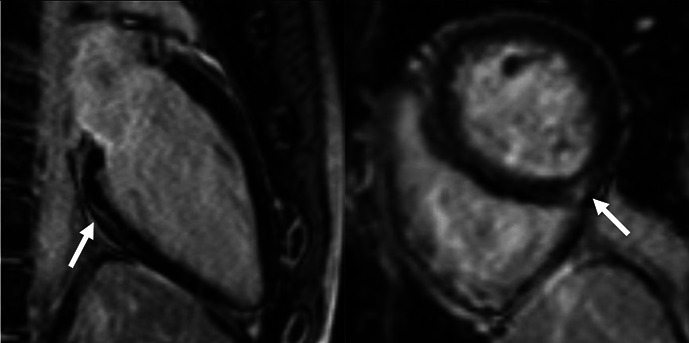
27 | 4 | LVEF: 61% GLS: −22% Septal native T1: normal Septal native T2: normal Pathological LGE/pattern: Yes—non-specific inferior and hinge point LGE T1 and T2 mapping value in the area corresponding with the LGE: 984 and 41 ms—normal |
|
Possible | Returned to sport, no persistent cardiac complaints at follow-up |
| 4. | Female | Long COVID-19
|
Troponin: normal 12-lead ECG: normal Holter ECG: normal Echocardiography: normal Exercise test (5 months after COVID-19 infection): normal |
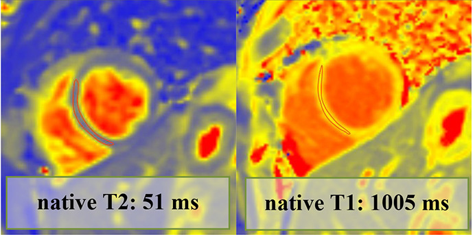
67 | <3 | LVEF: 67% GLS: −27% Septal native T1: grey zone normal/elevated Septal native T2: mildly elevated Pathological LGE/pattern: no |
|
Possible | Returned to sport, no persistent cardiac complaints at follow-up |
| 5. | Female | Moderate
|
Troponin: normal 12-lead ECG: PVC Holter ECG: trigeminy PVC on exertion Echocardiography: normal Exercise test (4 months after COVID-19 infection): normal |
19 | <3 | LVEF: 60% GLS: −22% Septal native T1: mildly elevated Septal native T2: mildly elevated Pathological LGE/pattern: No |
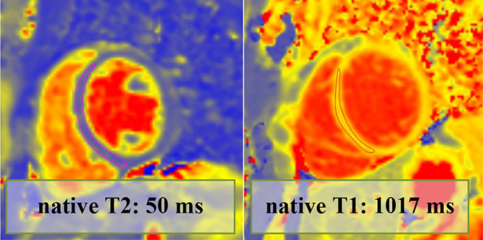
|
Possible | Returned to sport, no ongoing cardiac complaints. |
| 6. | Female | Mild
|
Troponin: elevated (hs troponin I: 28 ng/L—normal: <1.9 ng/L) 12-lead normal Holter ECG: NA Echocardiography: normal Exercise test: NA |
11 | <3 | LVEF: 55% GLS: −18% Septal native T1: mildly elevated Septal native T2: normal Pathological LGE/pattern: no |
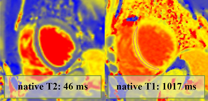
|
Possible | Returned to sport, no ongoing cardiac complaints |
| 7. | Male | Moderate
|
Troponin: elevated (hs troponin I: 225 ng/L—normal: <45 ng/L) 12-lead ECG: descending PQ segment depression Holter ECG: NA Echocardiography: decreased longitudinal strain, mild anterior and anteroseptal wall motion abnormality Holter ECG: NA |
120 | 10 | LVEF: 61% GLS: −20% Septal native T1: normal Septal native T2: normal Pathological LGE/pattern: Yes—pericardial involvement |
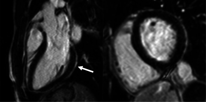
|
Possible | Returned to sport, no ongoing cardiac complaints |
CMR, cardiac magnetic resonance; GLS, global longitudinal strain; hsTnT, high-sensitivity troponin T; LVEF, left ventricular ejection fraction; NA, not applicable; PVC, premature ventricular complex.
