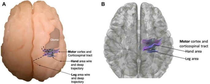
FIGURE 1.
Anatomic placement of glioma tumor near motor tracts. A, Superior view of the model showing the hand/arm and leg area wires exiting the superolateral surface of the brain at the precentral gyrus (motor cortex) and their deep trajectories corresponding with the corticospinal tracts. B, A 3D reconstruction of a magnetic resonance tractography used to guide wire placement posterior to the glioma in an anatomically accurate location.

