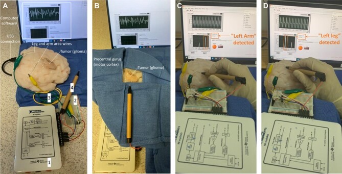
FIGURE 3.
Brain mapping simulator setup for data acquisition and general training and usage. A, Right superolateral view of brain mapping simulator shown with (1) NI myDAQ device, (2) custom circuit interfacing, (3) white matter tract models, and (4) handheld probe. Also depicted are the associated computer software interface and feedback mechanism (on screen connected through USB cable), locations of leg and arm white matter tract models in the simulator body, and location of glioma. B, Draped view of the brain mapping simulator showing a potential surgical training scenario with a glioma near the precentral gyrus (motor cortex) and the handheld electrical probe. C, Arm area stimulation. Surgeon uses handheld probe to stimulate the arm area in motor cortex and receives written and LED feedback on the screen (in orange). D, Leg area stimulation. Surgeon uses handheld probe to stimulate the leg area in motor cortex and receives written and LED feedback on the screen (in orange).

