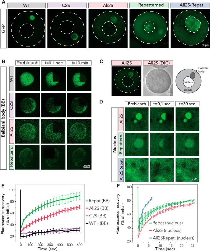
Figure 9.
Rationally designed Velo1 designs tune cellular localization and material state. (A) mRNAs encoding Velo1 designs and wild type Velo1 fragment F1 (Figure 8) fused to GFP were microinjected into stage I X. laevis oocytes. Oocytes were left to recover and express injected mRNAs overnight and imaged the next day. The cell membrane and nucleus are outlined in a white dashed line. (B) Internal rearrangement of fluorescent wild type or redesigned Velo1 (F1-GFP) particles after photobleaching in the Balbiani body. Note that Velo1Ali2S/Repat did not localize to the Balbiani body. (C) Overview, DIC image, and schematic of the oocyte with its Balbiani body in the Ali2S design. (D) Internal rearrangement of fluorescent redesigned Velo1 (F1-GFP) particles after photobleaching in the nucleus. Note that Velo1WT does not localize to the nucleus. (E) The fluorescent recoveries of the photobleached Velo1WT or redesigned Velo1 (F1-GFP) particles in Balbiani bodies in panel B and two other biological replicates were quantified over time. (F) The fluorescent recoveries of photobleached Velo1 designs in nuclei in panel C and two other biological replicates were quantified over time. For panels D and E, the fluorescence in the bleached region was quantified over time and normalized by an unbleached neighboring region. At least three oocytes per biological replicate were plotted. Scale bars are as indicated in the figure.

