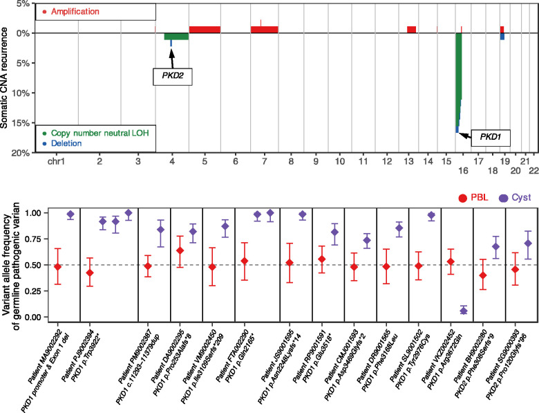
Figure 3.
Large-scale somatic structural alterations disrupt PKD1 and PKD2 in 90 cysts. Top panel: genome-wide patterns of large-scale somatic alterations in 90 cysts. The horizontal axis represents the genome. The vertical axis indicates the frequency of somatic copy number neutral LOH events (green) or deletions (blue), and chromosomal amplifications (red). Recurrently affected somatic regions where PKD1 and PKD2 genes reside are indicated. Bottom panel: shift in VAFs of germline pathogenic PKD1 and PKD2 variants in somatic LOH regions of cysts. In 17 cysts (purple) with somatic LOH events at PKD1 and PKD2 loci, the VAFs of the germline pathogenic variants shifted away from heterozygosity observed in matched PBLs (red). In all except one cyst, the LOH events led to increased VAFs of the pathogenic allele, suggesting biallelic inactivation of PKD1 or PKD2.

