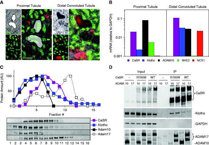
Figure 4.
Colocalization of the CaSR, Klotho, ADAM10, and ADAM17 in the kidney. (A) Laser capture microscopy of mouse kidney sections. Enlarged merged image of serial sections visualized by differential interference contrast (DIC) and immunofluorescence (IF) for DCT (specific sodium-chloride cotransporter; NCC in red) and PTs (lotus tetragonolobus agglutinin, LTA in green). (B) Quantitative PCR measurements of mRNA expression for DCT-specific NCX1 and PT-specific NHE3 with the CaSR, Klotho, and ADAM10. (C) Density gradient fractionation of mouse kidney extracts. Representation of the amount of protein as a function of gradient fraction number is shown. ADAM10 cofractionates with the CaSR and Klotho to a greater extent than ADAM17. (D) Coimmunoprecipitation of the CaSR, Klotho, and ADAM10 or ADAM17 from HEK-293 cells. HEK-293 cells or HEK-293 cells that stably express an HA-tagged CaSRWT or nonfunctional mutant CaSRR795W were transiently transfected with full-length Klotho and ADAM10 or ADAM17 (as indicated), extracted in RIPA buffer, and the lysates were incubated with anti-HA agarose. “Input” refers to cell lysates before immunoprecipitation; IP refers to the immunoprecipitated samples corresponding to the lysates in “Input”. The Input and IP samples were separated by SDS-PAGE, transferred onto a nitrocellulose membrane, and proteins were identified with specific antibodies as indicated on the right, except that ADAM10 and ADAM17 were identified with anti-HA antibodies. GAPDH, glyceraldehyde-3-phosphate dehydrogenase.

