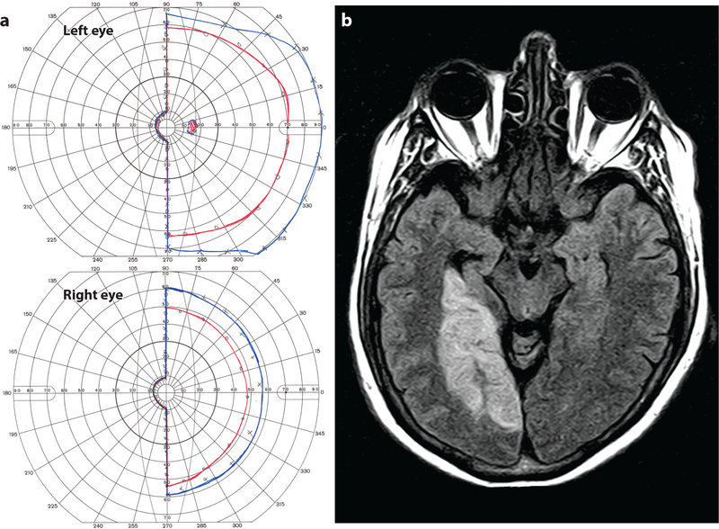
Figure 10.
Left hemianopia with macular sparing. (a) A 68-year-old man with left homonymous hemianopia and macular sparing; the I4e (red) and V4e (blue) isopters, mapped by Goldmann perimetry, are shown. (b) Axial FLAIR magnetic resonance (MR) image showing right posterior cerebral artery territory infarct with preservation of the occipital pole, presumably due to collateral flow from the middle cerebral artery.

