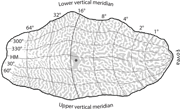
Figure 3.
Visual field map of striate cortex with polar coordinates from Horton & Hoyt (1991), scaled to a reconstruction of the pattern formed by ocular dominance columns in a human autopsy specimen (Adams et al. 2007). Rings of iso-eccentricity from the fovea are labeled dorsally, where they intersect the lower vertical meridian. The blind spot representation (*) is at the expected location, confirming the enormous cortical magnification of the macula. About half the cortex is devoted to the central 15°. Abbreviation: HM, horizontal meridian.

