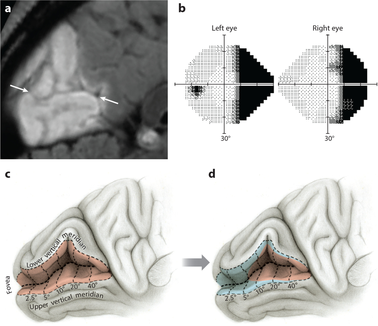
Figure 6.
Hemianopia with recovery along the vertical meridian is unphysiological. (a) A 52-year-old woman with infarction of the left occipital lobe from embolic occlusion of the posterior cerebral artery shown in a sagittal T2 FLAIR magnetic resonance (MR) image. Arrows mark calcarine fissure. (b) Humphrey 24–2 threshold tests performed 10 weeks later showing right homonymous hemianopia, with a strip of apparently intact right visual field approximately 5° in width along the vertical meridian. (c) Opened calcarine fissure showing the visual field map in left primary visual cortex (red shading). The vertical meridian is represented along the V1–V2 border; numbers denote degrees from the fovea. (d) Extent of cortical recovery (blue shading) required to produce the restored field along the vertical meridian mapped in panel b. There is no biological basis for such an eccentricity-dependent gradient in tissue recovery.

