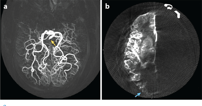
Figure 8.
3D digital subtraction angiogram reconstructions showing variation in perfusion of the occipital lobe by the posterior cerebral artery versus middle cerebral artery. (a) Maximum intensity projection after contrast agent injection into the basilar artery (yellow arrow). The posterior cerebral artery supplies tissue on the lateral convexity and, thus, all striate cortex. (b) A different patient, after injection into the right internal carotid artery, showing middle cerebral artery supply to the occipital pole (blue arrow), potentially supplying the macular representation. The right posterior cerebral artery territory is not filled, owing to congenital absence of the right posterior communicating artery. Images courtesy of Matthew Amans, M.D.

