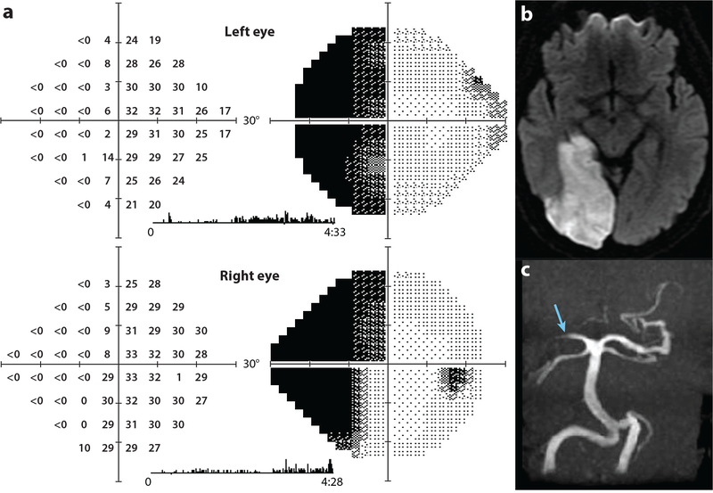
Figure 9.
Left hemianopia with macular splitting. (a) A 62-year-old man with left homonymous hemianopia tested by automated perimetry 4 months after stroke; thresholds measured in decibels (0 = 105 apostilbs) and corresponding grayscale representations are shown. The patient saw some stimuli along the vertical meridian on the blind side by sneaking saccades to the left. These saccades are captured by the eye monitor trace as vertical tic marks during the 4.5 minutes of testing. By confrontation testing the hemianopia was macular splitting. (b) Axial diffusion–weighted image showing acute right posterior cerebral artery infarction that encompassed the entire V1 by extending onto the lateral convexity of the occipital lobe. (c) Magnetic resonance (MR) angiogram showing occlusion of the right posterior cerebral artery (blue arrow).

