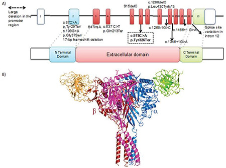
Figure 1.

A) 3-D structure of the SCNN1B protein (PDB 6BQN) black arrow indicates the mutated position on the protein. B) Close up view of the tyrosine aminoacid at the 326th position

A) 3-D structure of the SCNN1B protein (PDB 6BQN) black arrow indicates the mutated position on the protein. B) Close up view of the tyrosine aminoacid at the 326th position