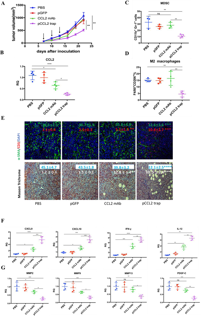
Figure 3.
pCCL2 trap outperformed CCL2 mAb and remodeled the immunosuppressive TME in 4T1 mouse model. A. Tumor inhibition effect of PBS, pGFP, pCCL2 trap and CCL2 mAb. 1×10⁶ 4T1 cells were orthotopically injected into mammary fat pad of six-weeks female BALB/c mice on day 0. Mice were randomly distributed into 4 groups on day 7 (n=5). Tumor volumes were recorded every other day by caliper measurement. Black arrow indicates the dosing schedule (50 μg pDNA or 200 μg mAb on day 7, 10 and 13). **p<0.01. B. The CCL2 mRNA expression in the tumor of mice received different treatments. Tumors were obtained at the end of the study (day 23, 10 days after the last treatment). *p<0.05, **p<0.01, ***p<0.001, ****p<0.0001. C-D. The quantitative analysis of MDSC and M2 macrophage population within TME on day 23. Cells were measured by flow cytometry. MDSC, myeloid-derived suppressor cells. E. Upper panel, immunofluorescence staining of tumor samples from different treatment groups using anti-α-SMA antibody (green) and anti-CD3-antibody (red). Cell nuclei were stained as blue using DAPI. Lower panel, collagen contents quantified by Masson Trichrome staining. Five random fields were chosen for statistical analysis in each treatment groups. Images were analyzed by Image J software and quantified with GraphPad 6.0. *p<0.05, **p<0.01, ***p<0.001, ****p<0.0001. Scale bar represents 100 μm. F. Relative mRNA expression of Th1 chemokines and cytokines in the tumor of mice received different treatments. Tumors were obtained at the end point of the study (day 23, 10 days after the last treatment). CXCL9: C-X-C Motif Chemokine Ligand 9; CXCL10: C-X-C Motif Chemokine Ligand 10; IL-12: interleukin-12; IFN-γ: interferon gamma. *p<0.05, **p<0.01, ***p<0.001. G. Relative mRNA expression of Th2 chemokines and cytokines in the tumor of mice received different treatments. Tumors were obtained at the end point of the study (day 23, 10 days after the last treatment). MMP, metallopeptidase. PDGF-C, platelet-derived growth factor C. *p<0.05, **p<0.01, ***p<0.001.

