
Figure 1.
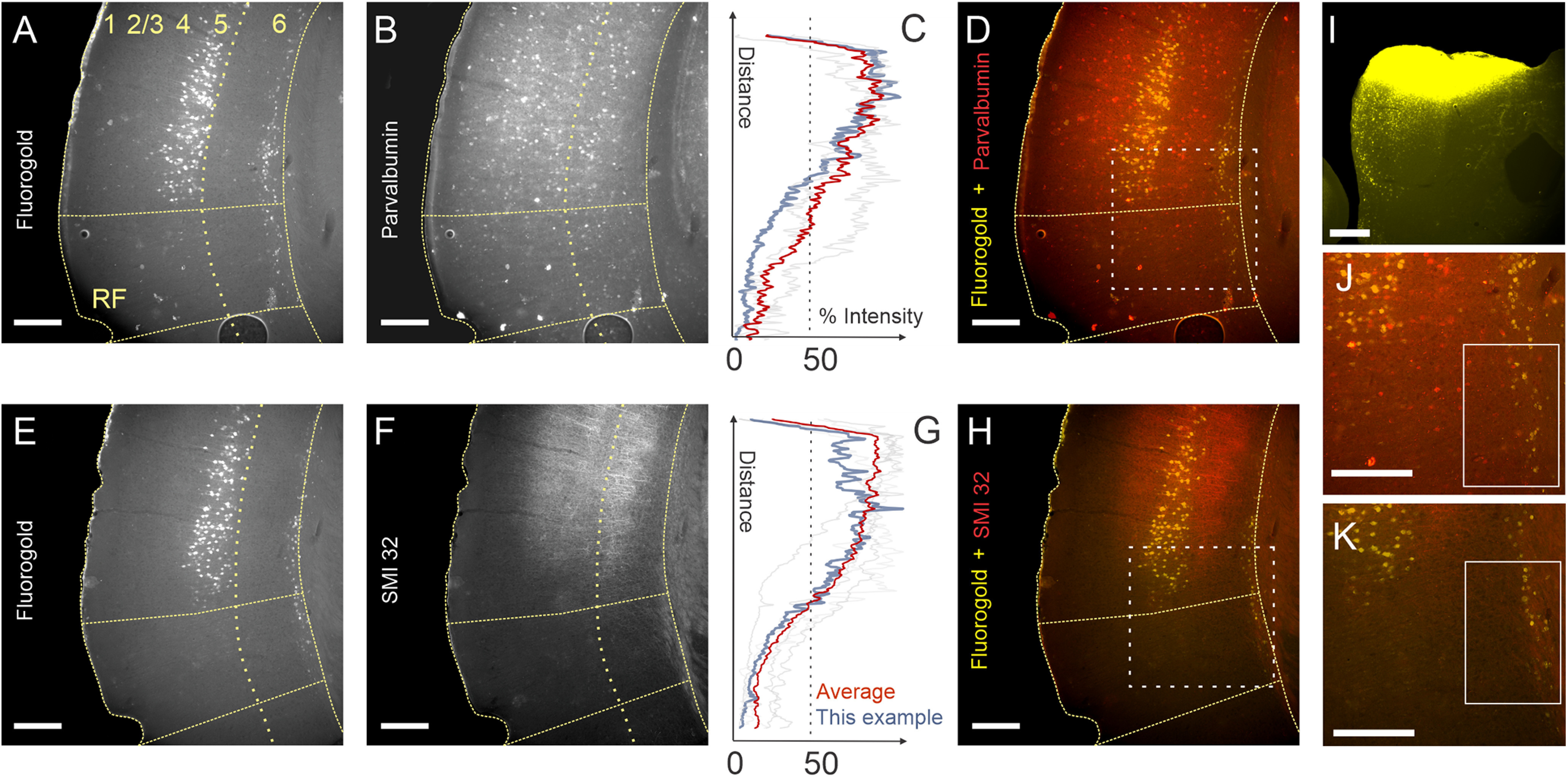
Partially segregated distributions of layer 5 and layer 6 corticocollicular neurons in mouse AC. A, E, Corticocollicular neurons in layers 5 and 6 from adjacent coronal sections from the same animal labeled with Fluoro-Gold after an injection into the IC. B, F, Parvalbumin (B) and SMI32 (F) immunoreactivity in the same sections. C, G, Fluorescence intensity profiles for PV (C) and SMI32 (G). Blue traces correspond to the intensity of the example mouse shown in this figure. The orange trace is the mean of all animals. Grayed-out traces are the individual other mice from this study. D, H, J, K, Layer 5 corticocollicular neurons are confined to PV- and SMI32-rich regions of the mouse AC (D, H), while many layer 6 corticocollicular neurons are found in PV– and SMI32– cortical regions (D, H, J, K). I, Corresponding injection of Fluoro-Gold in the left IC. Scale bar, 250 μm. Dotted boxes in D and H correspond to the areas shown in J and K, respectively. Solid boxes in J and K illustrate the presence of layer 6 corticocollicular cells without the presence of corresponding layer 5 corticocollicular cells.