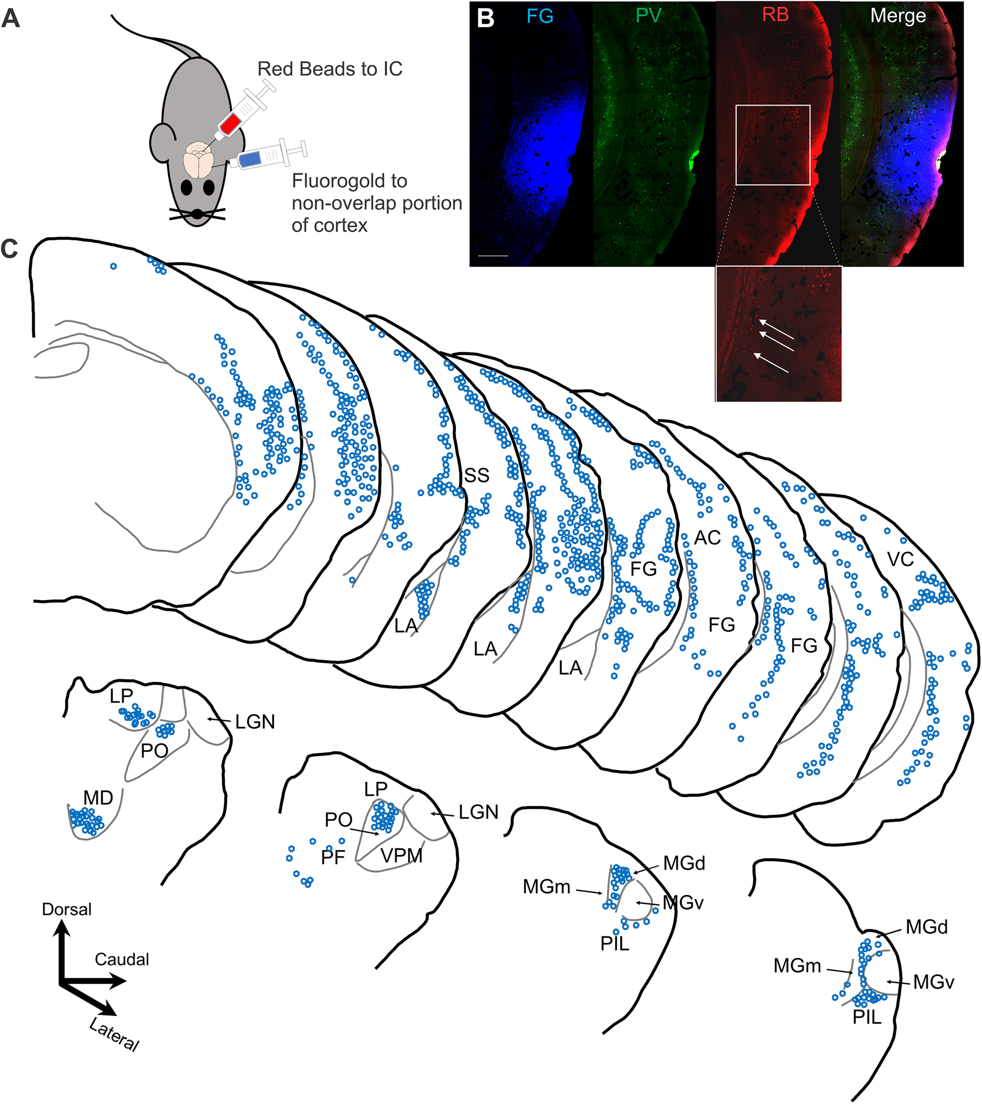
Figure 7.

Inputs to the cortical area containing layer 6 but not layer 5 corticocollicular neurons. A, Diagram of the dual-injection paradigm, with Fluoro-Gold being injected into the portion of the cortex containing a predominance of layer 6 corticocollicular cells and red retrobeads into the IC. B, The injection site showing Fluoro-Gold, the parvalbumin distributions showing diminished parvalbumin staining in the injection zone, and the red retrobead (RB) distribution. Box and expansion show the region of nonoverlap with layer 6 corticocollicular cells (arrows) without corresponding layer 5 corticocollicular cells. Scale bar, 500 μm. C, Top, The injection site (FG) and cortical areas where retrogradely labeled neurons were found. A significant portion of inputs came from the lateral nucleus of the LA, as well as somatosensory cortex (SS) and visual cortices (SS and VC, respectively). The bottom panel shows the distribution of neurons in the thalamus and associated structures. The majority of labeled neurons were found in the PO, LP, MD, PIL, and PF, the medial medial geniculate body (MGm), and the dorsal medial geniculate body (MGd), but not the lemniscal MGBv. For display purposes only, Fluoro-Gold is shown in blue.
