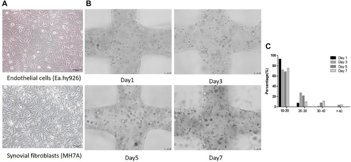
FIGURE 4.
Cellular morphological differences between 2D planar culture and 3D scaffolds. (A) EA.hy 926 and MH7A cells morphology in 2D planar culture. EA.hy 926 cells look epithelioid and MH7A cells are epithelioid and polygonal. Scale bar, 100 μm. (B) 3D Gelatin /alginate/EA.hy 926/MH7A scaffolds observed by a fluorescence microscope on culture days 1, 3, 5, and 7. Scale bar, 100 μm. Black arrows indicate cells and cellular spheroids in 3D scaffolds. (C) Distribution of spheroid diameter in 3D cell laden scaffolds on day 1, 3, 5, and 7.

