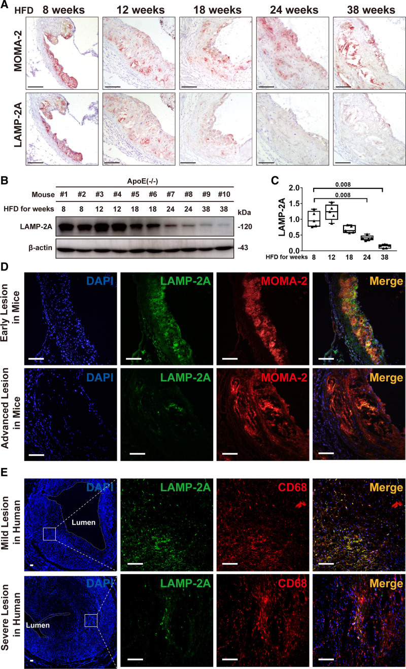
Figure 2.
Alterations in chaperone-mediated autophagy marker LAMP-2A (lysosome-associated membrane protein type 2A) in atherosclerotic lesions of mice and humans. A, Representative immunohistochemical images for detecting MOMA-2 (upper, specific for monocytes and macrophages) and LAMP-2A (lower) in 2 consecutive (upper and lower) frozen sections from the aortic root of ApoE−/− mice (male) fed a high-fat diet (HFD) for 8, 12, 18, 24, and 38 wk, respectively (n=5 in each group). The positive reactions of tissue sections were displayed as red color. Scale bar=100 μm. B, Representative Western blot analysis of LAMP-2A in whole-aorta lysates (including the aortic root) from ApoE−/− mice (male) fed an HFD for 8, 12, 18, 24, and 38 wk, respectively (n=5 in each group). C, Quantitative analysis of western blot analysis in 5 groups of mice. Five independent experiments were performed. Data were presented as medians and quartiles. Mice fed with HFD for 12, 18, 24, and 38 wk were compared with those fed with HFD for 8 wk, respectively. Statistical analysis was conducted using Kruskal-Wallis 1-way ANOVA with Nemenyi post hoc test. D, Double immunofluorescence analysis for detecting LAMP-2A (green particles) and MOMA-2 (red particles) in early lesions in mice (male) fed an HFD for 8 wk and in advanced lesions in mice (male) fed an HFD for 24 wk. n=5 in each group. Scale bar=100 μm. E Double immunofluorescence analysis for detecting LAMP-2A (green particles) and CD68 (red particles) in mild lesions (area stenosis ≤30%) and severe lesions (area stenosis ≥ 90%) in human coronary atherosclerotic plaques. n=5 in each group. Scale bar=100 μm.

