Figure 7.
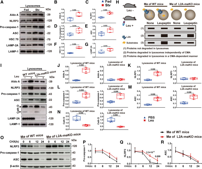
Deficient chaperone-mediated autophagy (CMA) inhibited degradation of the NLRP3 (NLR [NOD-like receptor] family, pyrin domain containing 3) inflammasome. A–G, Representative immunoblot images and quantitative analysis to detect the indicated proteins in lysosomes isolated from the peritoneal macrophages (MØs) of fed or starved (Stv) C57BL/6 mice (n=5, male). C57BL/6 mice were fed or starved for 24 h and were intraperitoneally injected with lipopolysaccharide (LPS) 4 h before MØ isolation to activate NLRP3 inflammasome. Five independent experiments were performed. Data were presented as medians and quartiles. Statistical analysis was conducted using Mann-Whitney test. H, Schematic diagram showing the method used to determine whether a protein is degraded by CMA: (1) proteins not degraded in lysosomes, (2) proteins degraded in lysosomes independently of CMA, and (3) proteins degraded in lysosomes through CMA. I–N, Representative immunoblot images and quantitative analysis of lysosomes isolated from the peritoneal MØs of wild-type (WT) and L2A-mØKO mice (macrophage [MØ-specific] LAMP-2A [lysosome associated membrane protein type 2A; L2A]-knockout mice) (n=5, male) who were starved for 24 h and then injected with or without leupeptin (Leu) 2 h before isolation. Aldo A (aldolase), a well-characterized CMA substrate, was used as a positive control. Five independent experiments were performed. Data were presented as medians and quartiles. Comparison was made only between PBS and Leu groups. Statistical analysis was performed using Mann-Whitney test. O–R, Representative immunoblot images and quantitative analysis of protein expression in peritoneal MØ extracts from WT and L2A-mØKO mice (n=5, male). MØs were treated with LPS for 6 h, and after LPS was removed, cells were treated with cycloheximide (CHX, 5 μg/mL) for various durations. Five independent experiments were performed. Data were presented as medians and quartiles. Comparison was made only between WT and L2A-møKO groups. Statistical analysis was carried out using Multiple linear mixed-effects modeling. ASC indicates apoptosis-associated speck-like protein containing a CARD (C-terminal caspase-recruitment domain); HSC70, heat shock cognate 71 kDa protein; and LAMP-2A (lysosome-associated membrane protein type 2A.

