
Figure 7.
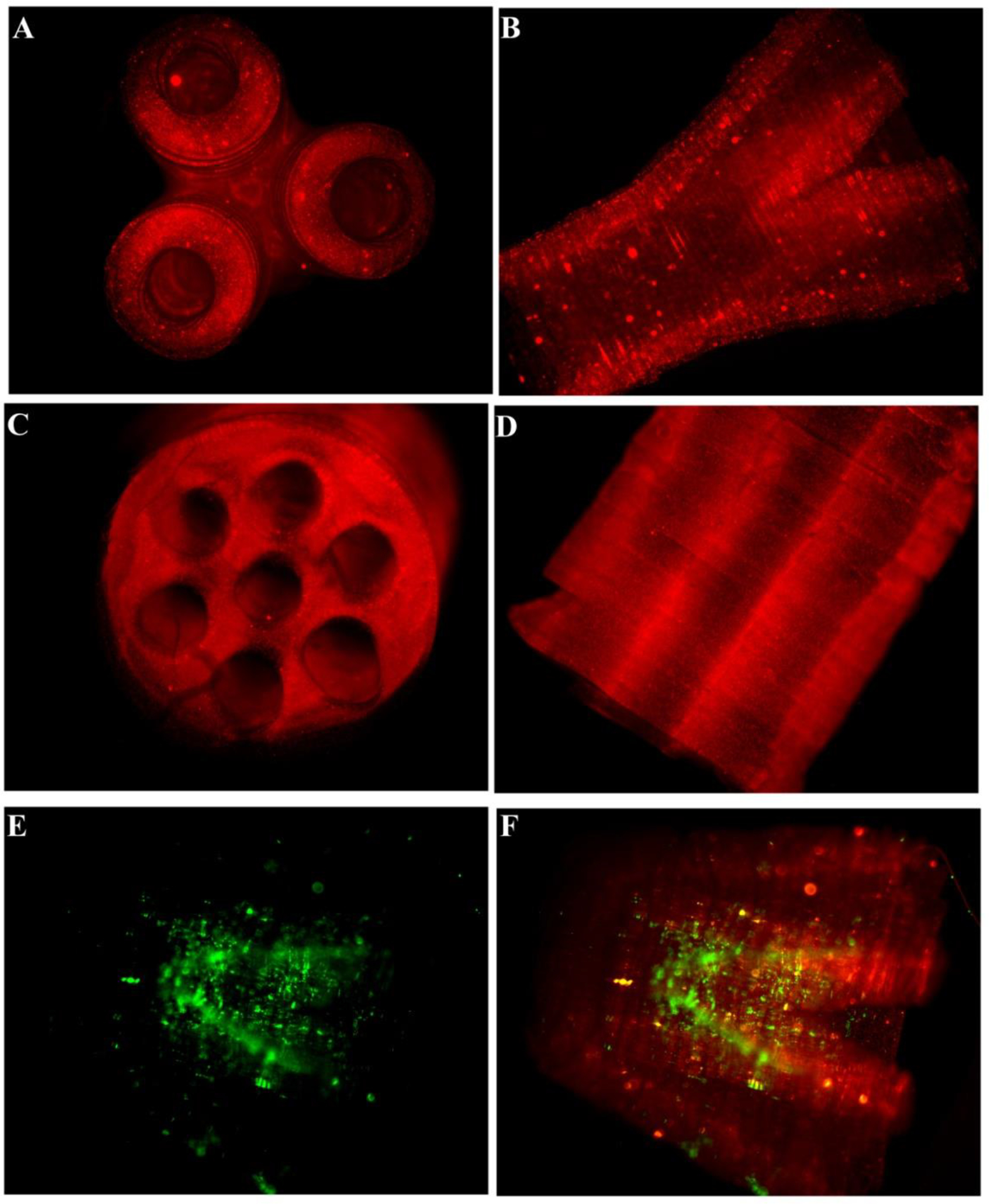
Fluorescence micrographs of the fabricated scaffolds. (A) and (B) demonstrate the lateral and top view of the branched scaffold. The scaffold starts as a single lumen conduit and divides into three branches at the other end. (C) and (D) demonstrate the lateral and top views, respectively, of the multi-lumen scaffold. (E) and (F) show the Schwann cells seeded inside the scaffold. The cells remained viable after 24 hrs when stained with calcein. Red fluorescent particles were added in the prepolymer solution to better visualize the scaffolds and the internal structure post-fabrication. (Scale bar, 1 mm)