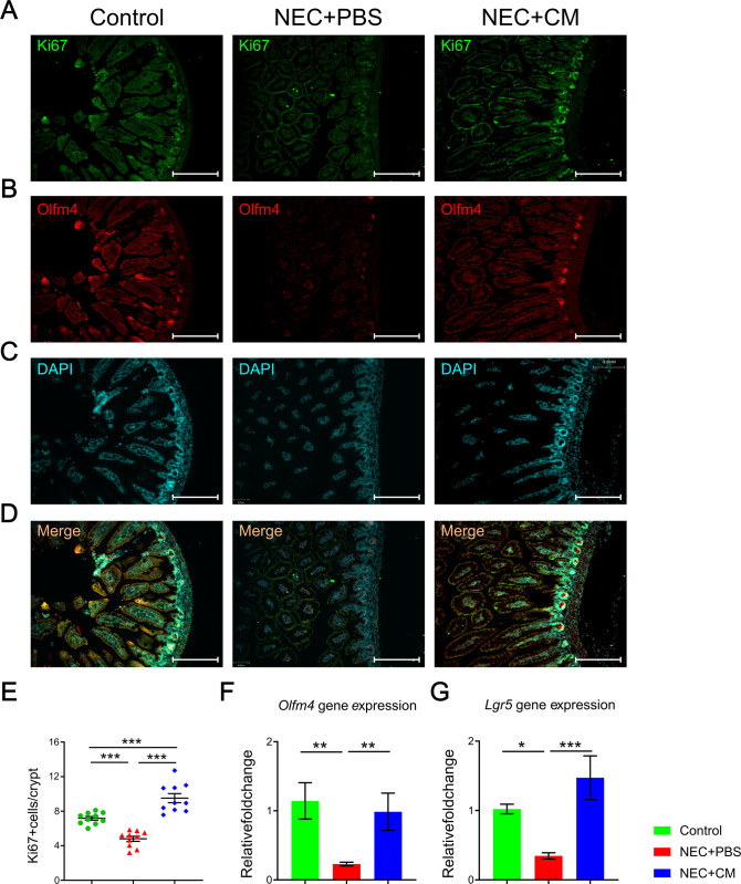
Fig 3. Intestinal proliferation and regeneration.
Representative immunofluorescent histomicrographs of the terminal ileum from the control, NEC+PBS, and NEC+CM groups, showing expression of (A) KI67, (B) OLFM4, (C) DAPI staining denoting the nuclei for each respective group, and (D) merging the 3 stains with yellow staining denoting positive KI67 and DAPI overlap. Relative quantitative expression for (E) KI67, (F) Olfm4, and (G) Lgr5 in the 3 groups. Data are presented as mean ± standard error, with significance of group comparisons based on one-way ANOVA and Tukey post-hoc tests. n = 10 for each group, *p<0.05, **p<0.01, and ***p<0.001.

