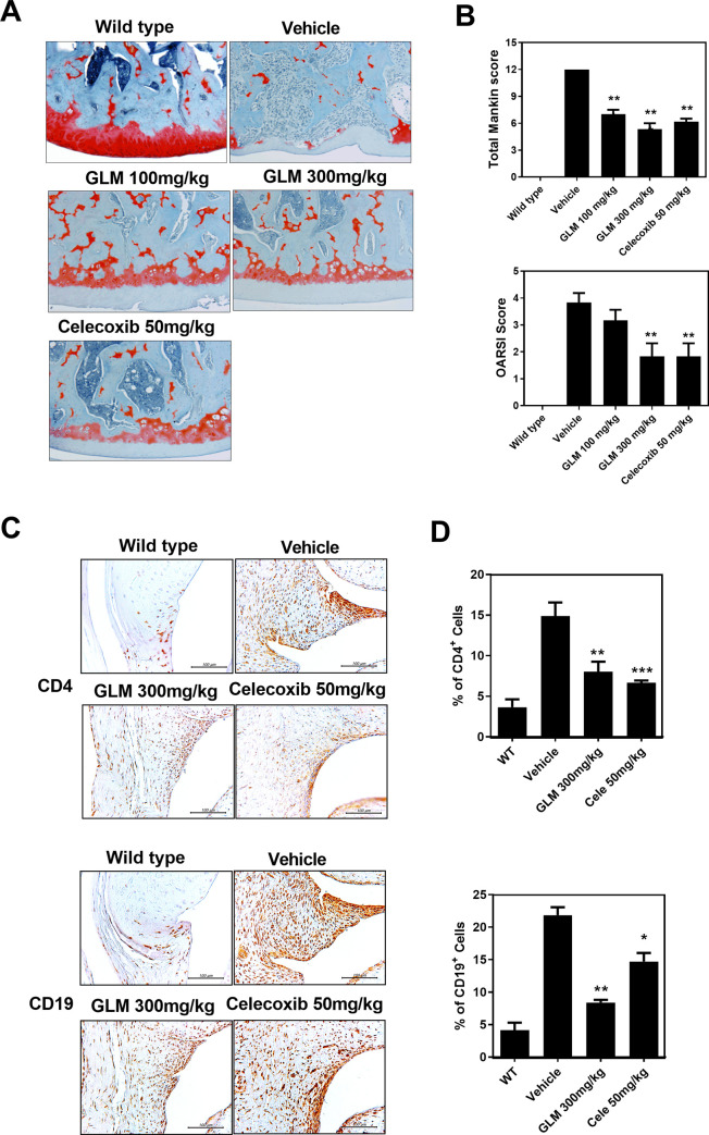
Fig 2. Histological evaluation of joints after oral administration of GLME to rats with MIA-induced OA.
Rats were injected with 3 mg of monosodium iodoacetate (MIA) (into the right knee). GLME was given orally every day from day 3 after MIA injection. The joints were resected on day 21 after MIA injection. (A) Knee joints from the OA rats. Joint samples were acquired from the WT, vehicle, GLM (100 mg/kg, 300 mg/kg) and Celecoxib (50 mg/kg) groups and stained with hematoxylin and eosin (original magnification x 200). (B) The OA lesions were graded on a scale of 0–13 using the modified Mankin scoring system that evaluates structure, cellular abnormalities, and matrix staining. *P < 0.05, **P < 0.01, and ***P < 0.001 compared to the MIA-injection group. (C) CD4 and CD19 expression levels in the synovia of OA rats as revealed immunohistochemically 21 days after MIA injection. Immunohistochemistry was used to evaluate representative sections of joints from MIA-injected rats given GLM, celecoxib, or the vehicle. Positive cells stain brown; the nuclei were counterstained with hematoxylin. The bar graphs represent the means ± SDs of the numbers of stained cells. *P < 0.05, **P < 0.01 compared to the MIA-injection group.

