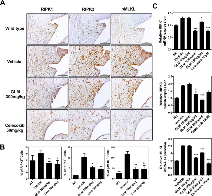
Fig 5. Reduced levels of necroptosis-related markers in OA rats and human chondrocytes.
(A, B) Representative immunohistochemical staining of RIP1, RIP3, and pMLKL in the synovia of non-OA rats (WT), vehicle-treated MIA-induced OA rats, and GLME-treated MIA-induced OA rats. (C) The levels of mRNAs encoding necroptotic marker genes (as revealed by real-time PCR) in human OA chondrocytes treated with IL-1β (20 ng/mL) and then co-cultured with GLME or celecoxib. *P < 0.05, **P < 0.01, and ***P < 0.001 compared to the vehicle-treated group.

