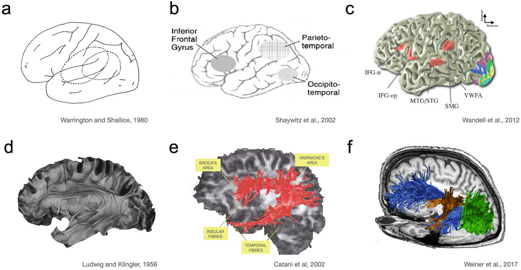
Fig. 1.
History of the reading circuitry. a Neurologists at the turn of the 20th century debated the location of the brain’s visual word form center, which was initially placed in the angular gyrus (Déjerine 1891). This debate played out through the early days of PET until a part of VOTC was discovered as the location of the “visual word form area”. The location was first confirmed by neurologists (Warrington and Shallice 1980). This debate was at the spatial scale of lobes. b Within the first decade of fMRI the three main components of the reading circuitry were defined as left inferior frontal, inferior parietal and occipitotemporal cortex (Pugh et al. 1996; Shaywitz et al. 2002). This early model continues to be influential and outlines the circuitry at the spatial scale of ~4cm (general locations within a lobe). c Over the next decade a more nuanced understanding of these regions emerged. Regions were precisely defined relative to sulcal landmarks at the millimeter spatial scale. Language-related regions are in red, while the other colors are used to illustrate retinotopic maps in visual cortex (Wandell et al. 2012). d, e, f A similar historical progression of anatomical precision for white matter pathways. d Ludwig and Klingler’s brain model provides a representation of the white matter tracts. Using Klingler’s method of dissecting the human brain after freezing it, these models revealed that axonal connections form large bundles, or fascicles (Ludwig and Klingler 1956). e Early diffusion tensor imaging based tractography showing a major white matter pathway connecting regions involved in language processing that is also crucial for reading. These in-vivo white matter tract reconstructions corroborated previous post mortem anatomical findings (Catani et al. 2002). f Recent advances in diffusion MRI allow for fine-grained representation of white matter pathways in relation to functionally defined regions within individual brains (Yeatman et al. 2012a; Takemura et al. 2015; Weiner et al. 2017b). The current state of the art has led to predictions of functional responses in individual brains, with millimeter precision, based on diffusion MRI measures of an individual’s white matter anatomy (Saygin et al. 2012; Grotheer et al. 2021). In each row there is a gradual increase of precision and spatial resolution, which became possible with the accumulation of knowledge from different modalities and the concomitant improvement in imaging technologies. Our current understanding builds upon these previous models and several aspects of the reading circuit are still being investigated in this rapidly evolving field of research. Copyrights: a Warrington EK, Shallice T. Word-form dyslexia. Brain 1980, Vol 103, 99–112, by permission of Oxford University Press. b Reprinted from Biological Psychiatry, Vol 52, Shaywitz BA, Shaywitz SE, Pugh KR et al., Disruption of posterior brain systems for reading in children with developmental dyslexia, 101-110, Copyright (2002), with permission from Elsevier. c Modified with permission from the Annual Review of Psychology, Volume 63 © 2012 by Annual Reviews, http://www.annualreviews.org e Reprinted from NeuroImage, Vol 17, Catani M, Howard RJ, Pajevic S, Jones DK, Virtual in vivo interactive dissection of white matter fasciculi in the human brain, 77–94, Copyright (2002), with permission from Elsevier. f Reprinted from Cortex, Vol 97, Weiner KS, Yeatman JD, Wandell BA, The posterior arcuate fasciculus and the vertical occipital fasciculus, 274-276, Copyright (2017), with permission from Elsevier.

