Fig. 3.
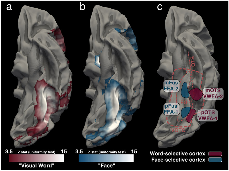
Word- and face-selective cortex (a,b) A meta-analysis conducted with Neurosynth of MNI coordinates associated with “Visual Word” (panel a) and “Face” (panel b). Data from (a) 117 and (b) 896 fMRI studies are shown on a group average template. When data are smoothed and analyzed on the MNI template a large portion of VOTC appears to be word-selective. In template space, word-selective and face-selective regions overlap covering much of the fusiform gyrus (FG) and the occipitotemporal sulcus (OTS). This type of group analysis obscures the anatomical organization of word-selective and face-selective cortex. (c) A schematic of the typical layout of word-selective and face-selective regions defined for an individual subject and shown on the fsaverage cortical surface. Word-selective regions can be reliably localized on an individual’s cortical surface and are spatially distinct from face-selective regions. Moreover, a sequence of at least 2 separate word- and face-selective regions can be identified within VOTC (Yeatman and White 2021). Anatomical boundaries of VOTC are shown as a thick dashed line and the FG and OTS are marked as thin dashed lines. Different naming schemes have been used to label these regions. Some groups use an anatomy-function labeling scheme where each region is named based on its anatomical location - posterior fusiform (pFus), mid fusiform (mFus), posterior OTS (pOTS), mid OTS (mOTS) - followed by its functional selectivity (e.g., words; faces) (Grill-Spector and Weiner 2014; Stigliani et al. 2015). Other groups have chosen to keep the widely used terms of “Fusiform Face Area (FFA)” and “Visual Word Form Area (VWFA)” and designated the subregions as FFA-1, FFA-2, VWFA-1, VWFA-2 (White et al. 2019b, a; Yeatman and White 2021). These two naming conventions refer to the same sub-regions and the catch-all terms “word-selective cortex” and “face-selective cortex” are used to refer to the collection of cortical regions that are selective for a given category.

